About Throat
The term throat refers to the entire front region of the neck. Vertically, it stretches from the bottom of the jaw down to the top of the clavicle, which is also referred to as the collarbone. Horizontally, it begins from the Adam’s apple to the cervical vertebrae which host the spinal cord. [1]
This small region of the neck is host to numerous organs which perform different functions, all crucial to a person’s survival. To begin with, let’s look at the areas which constitute the throat:
Throat anatomy (Gross)
The throat can be divided into 3 main parts: [2]
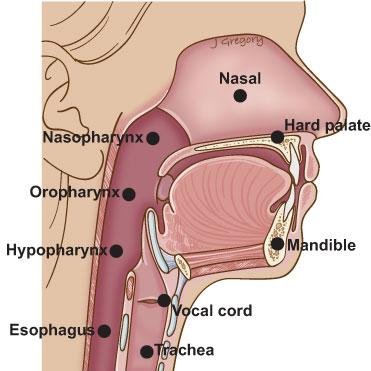
Oropharynx
This is the uppermost part of the throat that begins from the base of the tongue, which is a third of the tongue, through the oropharyngeal inlet, the tonsils, down to the vallecular, which is the space between the tongue and epiglottis. This area is responsible for receiving food and rolling it into a bolus for easier swallowing.
Hypopharynx
This is the area that contains the epiglottis as well as other forms of cartilage which make up the entrances to the esophagus and trachea. These cartilages ensure that the borders between these two organs are well defined while also contracting and relaxing to allow the smooth passage of food.
Larynx
At the bottom of the throat is the larynx which includes the vocal cords and other muscles responsible for speech.
Primary organs found in the throat
The throat may be a small part relative to the rest of the body, but it houses some very important organs: [3]
Vocal cords
The vocal cords are made up of tissue and stretch horizontally across the larynx. The vocal cords themselves only have a little muscle, but they are attached to muscles at the end that control the tightening of the vocal cords. In men, the vocal cords are thicker to facilitate the lower pitch, while women have a higher pitch attributed to the thinner vocal cords.
Besides speech, the vocal cords are responsible for the cough reflex at the first sign of food entrance, at which point they close up and initiate a cough to expel the food from the larynx.
Thyroid gland
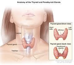
This organ is a butterfly-shaped organ found in the lower part of the throat below the Adam’s apple and is symmetrically positioned in the middle of the throat. Its main function is to control the rate of the body’s metabolism by producing thyroxine (T4) and triiodothyronine (T3) hormones after metabolizing iodine. The release of these hormones is regulated by thyroid stimulating hormone (TSH) which is produced by the pituitary gland at the behest of the hypothalamus.
When the hypothalamus detects an increased demand for energy, such as during a physically exerting exercise, it stimulates the pituitary gland to produce more TSH which subsequently stimulates the thyroid to produce more T3 and T4 hormones. These hormones increase the cells’ metabolic rate and they ‘burn’ more glucose to produce more energy. When there is no longer a need for that much energy, the hypothalamus reverses the process by lowering the production of TSH by the pituitary gland.
Epiglottis
This is just a flap made from elastic cartilage, but it serves a very important function in the throat – controlling the passage of air vs. solids and liquids. It is found between the esophagus and the trachea and closes the entry to the trachea during swallowing to prevent solid food and drink from entering the lungs.
The normal posture of the epiglottis is upright, where it remains vertical to allow air into the trachea and the lungs. It remains predominantly upright since an individual spends most of the time breathing rather than eating. When swallowing, the epiglottis folds into a horizontal position to cover the entry of the trachea and force the food down the esophagus. A covering of mucous membrane allows the food to slide down the throat more easily.
In case this process fails or doesn’t function properly, a secondary gag reflex is instituted, which is simply a contraction of the throat to prevent entry into the trachea. Furthermore, there are taste buds on the epiglottis, which also contribute to the taste of food.
Tonsils
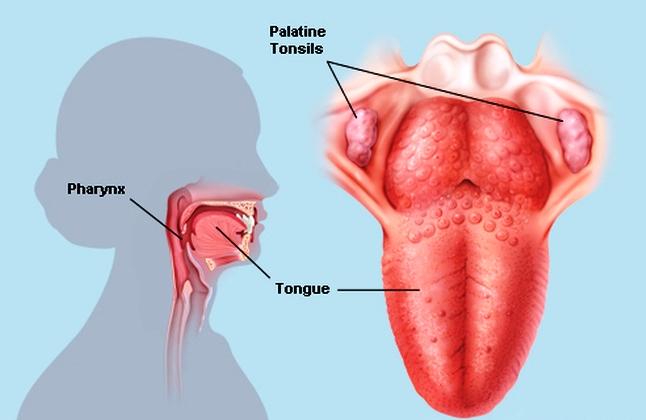
These are the tissues that can be visibly seen from the front of the mouth at the back of the throat. In fact, they are situated at both the back and the sides of the throat. Some people think the hanging mass is part of the tonsils, but that is just the uvula, responsible for producing saliva to keep the throat lubricated and also for speech.
The tonsils are a lymphoid tissue, instead, and their function is to fight off harmful pathogens from entering the throat. This explains why the tonsils become inflamed when they have been infected. The inflammation usually goes away using anti-inflammatory medications, but if the inflammation is too severe that it inhibits the individual from swallowing, it can be removed through surgery. This procedure is called a tonsillectomy.
Supportive structures of the throat
The throat would not function without other supportive structures, and these include:
Anatomy of the Throat: Blood Supply, Nerve Supply and Lymphatic System
Muscle
There are multiple groups of muscles around and within the throat all the way down from the oropharynx to the larynx. These muscles facilitate motion within the throat, allowing an individual to twist their neck and they also aid in swallowing and speech.
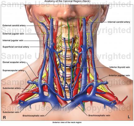
Blood supply
All the muscles in the throat need blood in order to function, and luckily, the throat gets a lot of blood from one of the largest arteries in the body, the carotid artery, which also supplies blood to the brain. The arteries pass through the throat on their way to the mouth to supply blood to the tongue and other parts of the mouth. This blood then drains into the jugular vein, another major vein, and there is a lot of communication between the blood vessels around the throat.
Nerve supply
The vagus nerve provides motor and sensory signals to the throat, all of which are essential for any normally functioning part of the body. These nerves are responsible for the enabling of all the above organs and ensure they function properly.
Lymphatic system
The lymphatic system is responsible for draining excess fluid from the circulatory system, and there are several lymphatic nodes in the neck that perform that function as well as protect the body from pathogens.
REFERENCE:
- Throat Anatomy Available from: http://www.emedicine.medscape.com/article/1899345-overview
- Anatomy of the Throat and Esophagus Available from: http://www.study.com/academy/lesson/anatomy-of-the-throat-and-esophagus.html
- Throat Anatomy: Throat Parts, Pictures, Functions Available from: http://www.diseasepictures.com/throat-anatomy-throat-parts-pictures-functions/
- Respiratory System Available from: http://www.innerbody.com/anatomy/respiratory