The word “foramen” means an opening or orifice. There are several foramina in the skull in which arteries, veins, nerves, ligaments, and muscles pass through. The foramen lacerum is one of these openings and it allows several structures of the body to pass through it [1, 2].
Anatomy of Foramen Lacerum
The foramen lacerum is the triangular orifice that is found in the middle cranial fossa. It is located anteromedial to the carotid canals in which the carotid arteries pass through. It has a fibrous covering that is being pierced by a small number of tiny vessels. This structure fills cartilage after birth [1, 2, 3, 4, 5].
The structure that serves as the anterior border of the foramen is the body of the sphenoid bone at the junction of the pterygoid process and greater wing. The basilar part of the occipital bone forms its medial border and the petrous apex forms its posterior border [1, 2, 3]. Although its size varies from one person to another, this foramen is about 9 mm in length and about 7 mm in breadth [1]. Figure 1 shows the location of the foramen lacerum.
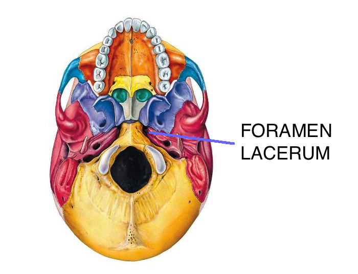
Figure 1- Foramen Lacerum
Clinical Significance
There are several structures that pass through the foramen lacerum.
Greater petrosal nerve
This nerve transits into the foramen lacerum before it joins the pterygoid canal nerve. The greater petrosal nerve branches from the facial nerve and is a part of the network of nerves that innervate the lacrimal gland. The main function of this nerve is to carry parasympathetic preganglionic fibers to the mucosal glands of the pharynx, palate and the nose [1, 2, 3, 4, 5].
Emissary vein
The emissary veins form a group of veins that serves as a connection between the intracranial cavernous sinus and the pterygoid plexus. These are the veins that drain the structures of the scalp, sinuses, face and pharynx. The emissary vein not only function as alternate drainage route of the intracranial venous system but it is also the structure that provides access to bacteria and other infectious agents to the cerebral veins and venous sinuses [1, 2, 3, 4, 5, 6].
Meningeal branch of the ascending pharyngeal artery
The ascending pharyngeal artery is a branch of the external carotid artery. It is the smallest branch of the carotid and it is located deep in the neck underneath the stylopharyngeus muscle and other branches of the carotid. Its main function is to provide adequate blood flow to the structures of the pharynx and the meningeal branch of this structure passes through the foramen lacerum [1, 2, 3, 4, 5].
Internal carotid artery
The internal carotid artery traverses partially through the foramen and the arteries and veins of pterygoid canal go through along with it. The main function of this branch of the common carotid artery is to supply the anterior portion of the brain, the eye, and the muscles of the eye. Branches of the internal carotid artery supply the forehead and the structures of the nose. It has several curvatures as it makes its way to the brain. The internal carotid artery looks like an italic letter “S” as it passes through the carotid canal and the side of the sphenoid bone body [1, 2, 3, 4, 5, 7].
Disorders of the Foramen Lacerum
Traumatic Lesion
The disorders of the foramen lacerum affect the structure that passes through this opening. A traumatic lesion that develops in this area may affect the cranial nerves that have a role in normal deglutition or swallowing. Although its occurrence may be rare, this can lead to more complications if not addressed accordingly. Treatment would require feeding thorough a nasogastric tube while an active and early rehabilitation of swallowing is being initiated. Recovery is possible even without doing any surgical intervention [8].
Nasopharyngeal carcinoma
The large or more aggressive variation of nasopharyngeal carcinoma finds a route through this foramen to reach the cavernous sinus, clivus, and temporal bone. This type of spread of the nasopharyngeal carcinoma is termed as a perivascular spread. Imaging tests will show that the structures of the foramen lacerum may be destroyed or eroded which suggests the aggressive nature of this process [9, 10]. The cranial nerves are also being affected when the carcinoma spreads in this method [5].
References
- Junior Dentist. (2012, August 14). Structures Passing through Foramina of Skull. Retrieved from Junior Dentist: http://www.juniordentist.com/structures-passing-through-foramina-of-skull.html
- Hacking, C., & Mudgal, P. (2005). Foramen Lacerum. Retrieved from Radiopaedia: https://radiopaedia.org/articles/foramen-lacerum
- Shrestha, S. (2011, June 2). Structures passing through foramina of the skull. Retrieved from Medchrome: http://medchrome.com/basic-science/anatomy/structures-passing-through-foramina-of-skull/
- Tauber, M., van Loveren, H., Jallo, G., Romano, A., & Keller, J. (1999). The enigmatic foramen lacerum. Neurosurgery, 386-391.
- Dentistry for Students. (2016). Anatomy and Structures passing through it. Retrieved from Dentistry for Students: http://www.studentistry.com/foramen-lacerum/
- Greenlee, J. E. (2010). Handbook of Clinical Neurology. Salt Lake City: Elsevier.
- Inner Body. (2017). Internal Carotid Artery. Retrieved from Inner Body: http://www.innerbody.com/anatomy/cardiovascular/internal-carotid-artery
- Lartigue, C., Roualdes, G., Muckenstum, B., Mesz, M., & Deglaire, B. (1987). [Deglutition disorders caused by an injury to the foramen lacerum posterior]. Annales Françaises d’Anesthésie et de Réanimation, 520-522.
- Knipe, H., & Gaillard, F. (2017). Nasopharyngeal carcinoma. Retrieved from Radiopaedia: https://radiopaedia.org/articles/nasopharyngeal-carcinoma
- Sham, J. S., Cheung, Y. C., Chan, F., & Leong, L. (1991). Nasopharyngeal Carcinoma: CT Evaluation of Patterns of Tumor Spread. American Journal of Neuroradiology, 265-270.