What is atrial myxoma?
Atrial myxoma is a benign tumour that is placed on heart walls. Due to its location however, it can induce all types of problems with blood flow and induce clotting and arrhythmias, which may induce severe symptoms in a person. Histologically this tumour consists of gelatinous tumour tissue. It is usually on a peduncle which is a couple of millimetres long.
The tumour may be between 1 and 15 cm in diameter. The tumour may be yellow or white, globule-shaped and in most cases is located in the left atrium (in 75% of people with myxoma), causing obstruction or a semi-obstruction of blood flow. If they are located in the right atrium they are sessile and solid, and cause fewer problems.
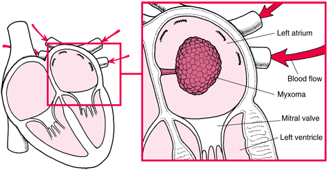
Figure 1 Position and appearance of myxoma in the left atrium (1)
Some myxomas appear sporadically, and some have genetical inheritance. This tumour doesn’t have a high prevalence, and occurs less frequently than secondary, metastatic tumours. According to studies, for some reasons, 75% of people with myxomas are women. Myxomas may produce some hormones and substances which may alter some body functions. (2)
Signs and symptoms
In first years as the tumour grows, the symptoms aren’t present, or are nonspecific. Depending on the size and location of the tumour, symptoms are different. If the right side of the heart is the site of myxoma, a person will experience swelling in the legs, abdominal swelling and fatigue, but these symptoms appear only with the severe right heart failure.
If it is located in the left atrium, the symptoms may be dizziness and syncope, heavy breathing, frequent respiratory infections, coughing, difficulties breathing when laying down without a pillow under head, but these too develop with the development of a heart failure. These symptoms appear because the blood flow is deprived and there is a damage of the structure and function of the valvulae.
Obstruction of blood flow and embolism are also common. Symptoms depend on the location of the myxoma. Myxoma may stay in the heart making an obstruction or may separate from the peduncle and travel through the bloodstream causing embolism somewhere else.
If the myxoma or a part of myxoma stop in the brain it will induce stroke or transient ischaemic attack. A person may suddenly feel loss of sensation to one side of the body, a headache, or faint.If the embolus travels to any organ it may deprive its blood circulation causing the ischaemia and possible necrosis and organ failure. Embolus may be very small and induce non-specific symptoms, or Raynaud syndrome (problem with circulation on hands), or lung hypertension or angina pectoris, etc. (3)
Diagnosis
Myxoma may induce similar auscultatory findings as mitral stenosis or regurgitation, endocarditis, or other diseases, which is why it is difficult diagnosing this tumour without ultrasound testing.
It is important to do the clinical evaluation, but the clinical picture will be different and non-specific in most of the cases. There are some findings that may appear on exam:
- Early diastolic sound due to physical presence of tumour
- Pathological sounds S3 and S4
- Systolic murmur if there is mitral regurgitation or diastolic rumble if there is a tricuspid regurgitation
- Loud S1 due to prolapse of tumour into the mitral orifice, imitating mitral stenosis clinical signs
- Clinical symptoms are: fever, coughing, weight loss, dizziness, fainting, haemoptysis (coughing blood), oedema, cyanosis, petechiae.
Valuable information are gathered from the ultrasound where it is visible that there is a mass in one of the cavities. The transesophageal approach brings more valuable and accurate information. MRI, CT, ECG and X ray findings contribute to other findings. (4)
Treatment
The treatment of myxoma isn’t effective with medications only, because there is a need for complete resection to relieve symptoms. If there is already a damage to the heart structure and function with signs and symptoms of a heart failure, medications are welcome to improve symptoms and delay the progression. Medical treatment includes:
- Antiarrhythmics,
- Treatment of heart failure (beta blockers or digoxin, oxygen therapy, ACE inhibitors, diuretics…),
- Anticoagulation therapy,
- Antibiotics for respiratory infections. (5)
Surgery
Definitive treatment option is surgical care. If the location is typical, i.e. in the left atrium the chance for recurrence is much lower than when it is atypically located.
The surgery is however, risky, for the tumour is easily fragmentated into peace, and there is a possibility of embolism during or after the surgery. This happens rarely. In most cases the success after surgery is favourable if the complete tumur was extracted. If some tissue was left, there is a chance of recurrence.
To assure the complete ressection, many surgeons use biatrial approach. tHe tumour is extracted with wide margins, and then, after surgery hystologically verified. If some of the tissue remained in the heart, there is a small chance of malignant alteration.
Works Cited
- Atrial myxoma. Cardiac health. [Online] [Cited: 3 11, 2017.] http://www.cardiachealth.org/ca-blog/atrial-myxoma.
- GK., Sharma. Atrial myxoma. emedicine Medscape. [Online] 2017. [Cited: 3 11, 2017.] http://emedicine.medscape.com/article/151362-overview.
- al., D Souza D et. Cardiac myxoma. Radiopaedia. [Online] [Cited: 3 11, 2017.] https://radiopaedia.org/articles/cardiac-myxoma.
- Atrial Myxoma: Trends in Management. . Lone RA, Ahanger AG, Singh S, et al. 2008, International Journal of Health Sciences. 2(2), pp. 141-51.
- Atral myxoma treatment. BMJ Best practise. [Online] [Cited: 3 11, 2017.] http://bestpractice.bmj.com/best-practice/monograph/1054/treatment/step-by-step.html.

